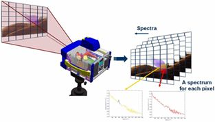
Spectral imaging technology is a new type of multi-dimensional information acquisition technology combining imaging technology and spectral technology, which can detect the data cube composed of two-dimensional spatial information and one-dimensional spectral information of the measured target, and obtain the spectral curves of different features after data processing.

Classification of spectral imaging technology

Spectral imaging technology originated in the 1980s, and its predecessor is multispectral remote sensing imaging technology. Due to the good information acquisition ability of spectral imaging, spectral imaging technology has been developed rapidly, and a variety of spectral imaging technologies have been developed, and imaging spectrometer products are constantly updated
.
There are various classification criteria for spectral imaging techniques, which can be divided into dispersive and interferometric spectral imaging techniques according to different spectroscopic methods. Both dispersive and interferometric spectral imaging techniques obtain two-dimensional spatial information and one-dimensional spectral information of the target by pushing or pendulum sweeping, which requires high stability of the platform and acquisition of spectral information of all spectral bands in the same exposure. The spectral imaging scheme using filters, whether using multiple filters to acquire image information of multiple wavelengths in parallel or using sequential filter switching, requires setting the appropriate exposure time according to the spectral response of the system so as to obtain the maximum signal-to-noise ratio.
Dispersive prism spectroscopy
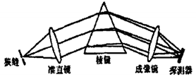
Dispersive prisms are the most commonly used and simplest spectroscopic imaging components, and the above figure shows a typical application of dispersive prisms in a spectral imager. As shown in the figure, the incident slit is located on the front focal plane of the collimation system. After the incident light passes through the collimation system, the slit is imaged on the focal plane detector by the imaging system according to wavelength through the prism.
Diffraction grating spectroscopy
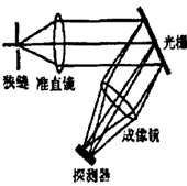
Diffraction grating is applied in the same way as a dispersive prism. The incident slit is located on the front focal plane of the collimation system, and after the incident light passes through the collimation system, the slit is imaged on the focal plane detector by wavelength through the grating.
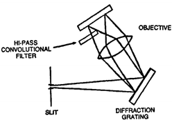
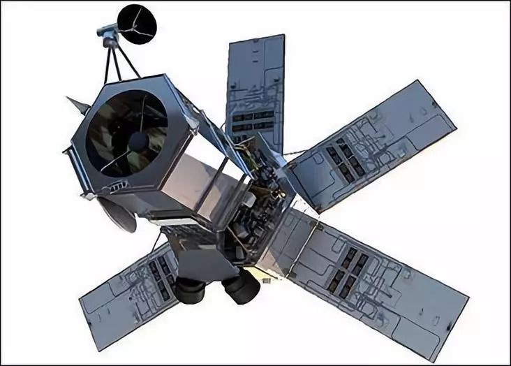
Another use of diffraction gratings is to place them in a dispersive beam, where light incident from the slit is directly incident on the grating without a collimation system, and a spectral virtual image of the target slit is obtained after diffraction by the grating, and the imaging system images the slit by wavelength at different locations on the surface array detector, an imaging technique that has been applied to the conceptual design of the OrbView-4 satellite’s tactical remote sensor.
The more mature international airborne and aerospace hit-in dispersive spectrometers are based on diffraction gratings, such as AVIRIS from the U.S. Jet Propulsion Laboratory, CASI from Canada, AISA from Finland, and instruments and equipment such as the spectroradiometer MODIS.
Binary spectral component spectral technology
The binary optical element is both a dispersive and imaging element, using a monochromatic surface array detector to scan the selected wavelength imaging range in the direction of the optical axis, with each position corresponding to the imaging area of the corresponding wavelength. The binary optical element concentrates the incident light like an ordinary lens, but it is based on the diffraction principle, where the effective focal length of the chromatic aberration produced by diffraction is inversely proportional to the wavelength.
Unlike prisms or grating elements that disperse along the direction perpendicular to the optical axis, binary optics disperse along the axis, and the spectral resolution of an imaging spectrometer with binary optics is determined by the size of the detector. This structure imaging spectrometer is compact and has high diffraction efficiency.
Acousto-optical tunable filter spectroscopy technology
Acousto-optic tunable filter (AOTF) is a new type of dispersive element, which consists of three parts: acousto-optic medium, transducer array and acoustic terminal. According to the principle of acousto-optic diffraction, when the complex color light is incident to the acousto-optic medium at a specific angle, the incident light satisfying the momentum matching condition is diffracted by the ultrasonic wave into two orthogonal monochromatic beams, which are located on both sides of the zero-level light, due to the acousto-optic interaction. Changing the frequency of RF signal, the wavelength of diffracted light also changes accordingly. Continuous and rapid change of the frequency of the RF signal can achieve fast spectral scanning in the wavelength range of the diffracted light.
Interferometric imaging spectrometer
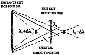
Since the spectral resolution of dispersive imaging spectrometer is inversely proportional to the width of the incident slit, to obtain higher spectral resolution, the width of the slit has to be continuously reduced, so that the luminous flux of the system is reduced, resulting in low detection sensitivity. As the technical specifications of imaging spectrometer requirements improve, especially in terms of spatial resolution, spectral resolution, and detection capability of weak signals, dispersive imaging spectrometers gradually fail to meet the requirements. Interferometric imaging spectrometer has the advantages of high spectral resolution and high energy utilization in principle, which can meet the increasing application requirements and gradually become a research hotspot in the field of imaging spectroscopy.
The main spectroscopic techniques of interferometric imaging spectrometer are Michelson interferometry, triangular common optical path interferometry, birefringent interferometry and so on. In recent years, the technique of using liquid crystal tunable filters to obtain polarized light and then interfering has been developed. In addition to the above two-beam interference techniques, there are also spectroscopy techniques based on multi-beam interference.
Filter-based imaging spectrometer
Filter-based imaging spectrometer is to add a filter to the optical path as a beam splitting element, by changing the filter to obtain different spectral channels. Filter-based spectral imaging changes the central wavelength by electrical tuning. The wavelength is adjusted once, the camera is exposed once, and the system records the two-dimensional image information of that wavelength, and then the next transmitted wavelength is set in. The cycle continues until the image acquisition task is completed for all wavelengths and the final spectral data cube is obtained.
Application of spectral imaging technology
Spectral imaging technology is a perfect combination of spectral analysis technology and image analysis technology, which has both spectral resolution capability and image resolution capability, allowing qualitative, quantitative and localization analysis of the object under test. Using the spectral differences of the surface components of the object, precise identification and localization of the target can be achieved, which has wide applications in the fields of material identification, remote sensing detection, medical diagnosis, etc.
The development of spectral imaging technology has gone through three stages: multispectral, hyperspectral and hyperspectral imaging. It is because imaging spectrometer can obtain multi-band image data with narrow band width, so it is mostly used for spectral analysis and identification of ground objects. With the continuous improvement of spectral resolution, the acquired target spectral information is more fine, and the applications in military, agriculture, medicine, resource exploration, geological investigation and other fields are becoming more and more extensive.
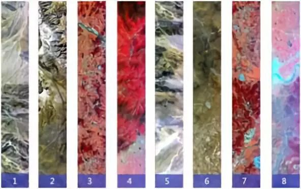
In the military, because of its ability to distinguish feature types spectrally, the imaging spectrometer is known as an important battlefield reconnaissance tool because of its strong advantages in fine classification of features, target detection and change detection head. Spectral images can distinguish between real and camouflaged targets in natural grass backgrounds and quickly detect small tactical targets in desert backgrounds.

In civil use, spectral imaging originated from the study of geological and mineral resources identification, especially the detection of special minerals such as mineralized alteration rocks, and gradually expanded to vegetation ecology, marine and coastal water color survey, water body detection, snow and ice, soil and atmospheric research. The spectral image with fine resolution has the characteristic of unity of map and spectrum, which can accurately detect the spectral information of vegetation growth characteristics, and even identify and estimate the chlorophyll, i.e., suspended matter content in waters, and detect the chemical pollution of water quality. Fine spectral imaging has become a hot spot for research at home and abroad, and scholars are using fine spectral imaging technology to quantify the material mechanism detection research at a more microscopic scale.